

An ECG (Electrocardiogram) is a medical test used to measure the electrical activity of the heart over a period of time. It records the electrical impulses or waves that trigger heartbeats, allowing doctors to monitor the heart’s rhythm, detect abnormalities, and identify potential issues like arrhythmias, heart attacks, or other cardiac conditions.
How it Works:
- The heart generates electrical signals that trigger muscle contractions, which pump blood.
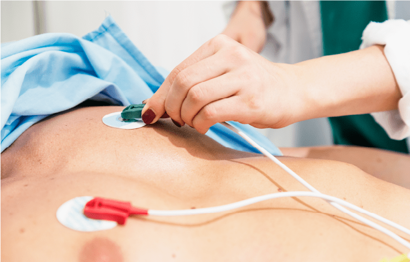
- An ECG records these electrical signals using electrodes placed on the skin, typically on the chest, arms, and legs.
- The signals are then displayed as waves on a graph, showing the heart’s rhythm and electrical activity.
2. The Waves:
The ECG graph typically shows several waves:
- P wave: Represents the electrical impulse from the atria (upper chambers of the heart).
- QRS complex: Reflects the electrical impulse as it moves through the ventricles (lower chambers of the heart), leading to contraction.
- T wave: Represents the recovery phase of the ventricles after contraction.
3. Purpose of an ECG:
- Diagnose heart problems: It helps detect irregularities in the heart’s rhythm (arrhythmias) or signs of heart attacks (myocardial infarctions).
- Monitor heart health: It can be used to track the progress of a heart condition or the effectiveness of treatments.
- Check heart rate: It provides the heart rate and rhythm, which are important indicators of cardiac function.
4. Types of ECG:
- Resting ECG: Done while the patient is at rest.
- Stress ECG: Performed while the patient is exercising (e.g., walking on a treadmill) to see how the heart responds to stress.
- Holter monitor: A portable ECG worn for 24-48 hours to track the heart’s activity over an extended period.
- Event monitor: Similar to a Holter monitor but used for a longer duration to capture occasional heart issues.
5. ECG Interpretation:
- The doctor will analyze the wave patterns to identify abnormalities in rhythm, structure, or electrical activity. For example:
- Arrhythmias: Irregular heartbeats, like atrial fibrillation.
- Heart attack: Specific changes in the waves can indicate recent or past heart attacks.
- Enlarged heart: Certain patterns can suggest heart chamber enlargement.
6. Preparation for an ECG:
- Typically, no special preparation is needed.
- The patient may be asked to lie down, and the skin where electrodes will be placed should be clean and free from oils or lotions.