

It seems like you might be referring to angiography, a medical imaging technique used to visualize the inside of blood vessels and organs. Angiography is most commonly used to check for blockages, aneurysms, or abnormalities in the blood vessels.
What is Angiography?
Angiography is an imaging technique that uses X-rays and a contrast dye to visualize the blood vessels in various parts of the body, like the brain, heart, kidneys, or lungs. The procedure helps doctors see any blockages, narrowing, or abnormalities in the blood vessels that might be affecting blood flow.
How Angiography Works:
Preparation:
- The patient is typically asked to fast for several hours before the procedure.
- A contrast dye (also called a “contrast agent”) is injected into the blood vessels through a catheter, which makes the vessels visible on the X-ray images.
- The catheter is usually inserted through a small incision in the groin or arm, where the doctor threads it into the relevant blood vessels, depending on what part of the body is being examined.
The Procedure:
- Once the catheter is in place, the contrast dye is injected. The dye travels through the blood vessels, and X-ray images are taken to capture detailed pictures of the blood vessels.
- The procedure typically takes 30 minutes to an hour, depending on the complexity of the case.
Types of Angiography:
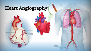
- Coronary Angiography: Used to visualize the blood vessels of the heart. It is often used to diagnose coronary artery disease (blockages in the heart’s arteries).
- Cerebral Angiography: Used to visualize the blood vessels in the brain, often to check for aneurysms, strokes, or other abnormalities.
- Pulmonary Angiography: Used to visualize the blood vessels in the lungs, particularly in the diagnosis of pulmonary embolism (a blockage in the lungs).
- Peripheral Angiography: Focuses on the blood vessels in the limbs and is often used to diagnose peripheral artery disease (PAD), which affects blood flow to the legs and feet.
- Renal Angiography: Visualizes the blood vessels of the kidneys and is used to check for conditions like kidney artery stenosis (narrowing of the arteries supplying the kidneys).
Post-Procedure:
- After the procedure, the patient may need to rest for a few hours, especially if the catheter was inserted through the groin.
- The contrast dye used in angiography is typically cleared from the body through the kidneys, so drinking fluids afterward is encouraged to help flush out the dye.
Uses of Angiography:
Detecting Blockages or Narrowing:
- It is commonly used to diagnose atherosclerosis (plaque buildup in the arteries), which can cause blockages that lead to heart attacks, strokes, or other complications.
Diagnosing Aneurysms:
- Angiography can detect aneurysms, which are abnormal bulges or weaknesses in blood vessel walls, which can rupture and cause severe bleeding.
Identifying Abnormal Blood Flow:
- It can reveal abnormal connections or growths between blood vessels, such as arteriovenous malformations (AVMs).
Evaluating Organ and Tissue Blood Flow:
- It is often used to assess blood flow in various organs, especially after trauma or surgery, to make sure that blood is circulating properly.
Guiding Treatment:
- Angiography is sometimes used during certain treatments, like angioplasty (a procedure to widen narrowed blood vessels) or stent placement to treat blockages.
Risks and Considerations:
While angiography is generally considered safe, there are a few risks to be aware of:
- Allergic Reaction to Contrast Dye: Some people may be allergic to the contrast dye used in angiography.
- Kidney Problems: The contrast dye can cause temporary kidney problems in some patients, especially those with pre-existing kidney conditions.
- Bleeding or Infection: As the procedure involves inserting a catheter, there is a small risk of bleeding or infection at the insertion site.
- Radiation Exposure: Angiography involves exposure to X-rays, but the levels are typically low and considered safe for most people.